FIP in Cats: Is It Contagious, How Is It Diagnosed, and Can It Be Cured?
What Is Feline Infectious Peritonitis (FIP)?
Few phrases strike panic into the heart of a cat parent quite like Feline Infectious Peritonitis (FIP). Historically considered a 100% fatal, untreatable systemic illness, FIP has long been one of the most feared, heartbreaking, and misunderstood diagnoses in veterinary medicine. When a veterinarian mentions the possibility of FIP, cat owners are often overwhelmed by intense grief, confusion, and fear about what lies ahead.
However, the medical narrative surrounding FIP has transformed dramatically over recent years. Thanks to extraordinary advancements in veterinary research and novel antiviral therapeutics, FIP is no longer an automatic death sentence. Today, it is a fully treatable condition with remarkably high rates of full, permanent recovery when diagnosed accurately and managed early in the disease process.

To fully understand how FIP develops, one must first understand the virus that precedes it: the Feline Coronavirus (FCoV). It is essential to recognize that feline coronavirus is extraordinarily common throughout the worldwide domestic cat population. In multi-cat environments, catteries, animal shelters, and rescue foster networks, up to 80% to 90% of cats test positive for antibodies to FCoV. Even in single-cat households, the lifetime exposure rate hovers between 20% and 40%.
In the vast majority of felines, this benign precursor—known as feline enteric coronavirus—resides completely harmlessly within the epithelial cells of the lower gastrointestinal tract. Most cats infected with benign FCoV experience either no clinical symptoms whatsoever or suffer only mild, self-limiting gastroenteritis, such as occasional soft stools, minor diarrhea, or brief bouts of vomiting that resolve on their own within a few days without aggressive medical intervention.
However, a tragic internal biological event occurs in approximately 5% to 10% of coronavirus-infected cats. Instead of remaining safely confined within the mucosal lining of the intestines, the benign enteric virus undergoes a random internal genetic mutation within the individual cat’s body. This critical mutation fundamentally alters the cell tropism of the virus, granting it the dangerous capability to infect and replicate inside white blood cells—specifically macrophages and monocytes.
Once inside these roving immune cells, the mutated virus hitches a ride throughout the cat’s entire circulatory system. The host’s immune system attempts to mount a massive response to destroy the invader, triggering severe, systemic, immune-mediated inflammation. This internal battle damages blood vessel walls (causing widespread vasculitis), allows protein-rich fluids to leak into body cavities, and forms inflammatory tumor-like nodules (granulomas) across multiple vital internal organs. This mutated, systemic hyper-inflammatory condition is what veterinarians officially diagnose as Feline Infectious Peritonitis.
How Do Cats Get FIP? Causes, Risk Factors, and Contagion Myths
A frequent question asked by heartbroken cat owners is: "Did my cat catch FIP from another cat at the shelter or clinic?" or "Is my other resident pet at immediate risk of catching FIP right now?"
The short, evidence-based answer is nuanced: FIP itself is generally NOT directly contagious from cat to cat. What IS highly contagious is the harmless, underlying Feline Enteric Coronavirus (FCoV).
How Feline Coronavirus Spreads
Understanding viral transmission requires separating the benign precursor virus from the internal mutated disease state:
- Fecal-Oral Transmission Route: FCoV is shed primarily in the feces of infected cats. Healthy cats ingest viral particles during normal daily activities—most commonly by sharing litter boxes, stepping in contaminated litter and subsequently grooming their paws, or sharing food and water bowls.
- Environmental Resilience: Microscopic viral particles can survive on carpet fibers, bedding, grooming brushes, clothing, and litter scoops for several weeks under ambient room temperatures.
- The Mutation Exception: When a cat develops FIP, the mutated virus is contained inside its internal macrophages, tissues, and fluids. The cat rarely sheds the mutated FIP strain in its feces. Therefore, if Cat A develops FIP, Cat B will not "catch" FIP directly from Cat A. However, Cat B may acquire the benign FCoV strain from the shared environment, which could theoretically mutate within Cat B if individual risk factors align.
Key Risk Factors That Trigger Viral Mutation
Why does the virus mutate in one cat while leaving nine housemates completely unaffected? While researchers continue to study specific genetic triggers, veterinary evidence points to a combination of age, immune status, stress, and genetic predisposition:
- Age Vulnerability: Over 70% of clinical FIP cases occur in young cats under two years of age, with peak incidence seen in kittens between 3 and 16 months old. Young animals possess immature immune systems that struggle to contain viral replication. Senior cats over 10 years old with waning immunity represent a secondary spike in FIP cases.
- Stress and Immunosuppression: Stress plays a massive physiological role in viral mutation. Major life events—such as adoption, rehoming, long-distance transport, spay or neuter surgery, recent vaccination, overcrowding, or concurrent parasitic infections—cause spikes in cortisol. Elevated cortisol suppresses cell-mediated immunity, giving benign gut viruses the operational window needed to mutate and spread.
- Purebred and Genetic Susceptibility: Certain pedigree breeds—including Bengals, Abyssinians, Ragdolls, Rexes, and British Shorthairs—demonstrate statistically higher rates of FIP. This suggests underlying hereditary immune vulnerabilities regarding how specific genetic lines respond to coronavirus challenges.
Unsure If Your Cat Is at Risk?
If your cat was recently adopted, underwent surgery, or is exhibiting unexplained lethargy and loss of appetite, immediate screening can make all the difference.
Recognizing FIP Symptoms: Wet FIP vs. Dry FIP
FIP presents in two main clinical forms: Wet (Effusive) FIP and Dry (Non-Effusive) FIP. In clinical practice, many cats also display a Mixed FIP profile, starting with dry lesions and eventually developing fluid build-up, or vice versa. Recognizing these signs early is the single most critical factor in achieving a complete cure.

1. Wet (Effusive) FIP Symptoms
Wet FIP accounts for approximately 60% to 70% of diagnosed cases. It is characterized by severe immune-mediated vasculitis, which causes protein-rich blood plasma to leak out of blood vessels and pool inside body cavities. Wet FIP progresses rapidly over days or weeks.
- Abdominal Swelling (Ascites): The classic clinical sign of Wet FIP is a noticeably distended, "pot-bellied" appearance. The cat’s abdomen expands with fluid while the surrounding skeletal muscle rapidly wastes away along the spine and hips. The belly feels firm yet fluid-filled upon gentle palpation.
- Pleural Effusion (Chest Fluid): When fluid accumulates inside the thoracic cavity, it compresses the lungs. This leads to respiratory distress, rapid shallow breathing, labored abdominal breathing, and open-mouth panting. This is a medical emergency.
- Refractory High Fever: Cats with Wet FIP display persistent or fluctuating high fevers (often between 103.5°F and 105°F / 39.7°C–40.5°C) that fail to respond to standard broad-spectrum antibiotics or short-term steroids.
- Rapid Weight Loss and Anorexia: Complete disinterest in food, sudden weight loss, severe lethargy, and a dry, unkempt coat.
2. Dry (Non-Effusive) FIP Symptoms
Dry FIP progresses more slowly over weeks or months. Because fluid does not accumulate in large quantities, diagnosis can be exceptionally challenging. Instead of systemic vessel leakage, the body forms localized, nodular inflammatory lesions (granulomas) on surface structures of internal organs, the eyes, or the central nervous system.
- Ocular Lesions (Eye FIP): Inflammation damages eye structures. Symptoms include sudden changes in iris color, cloudiness inside the cornea, keratic precipitates (clumps of inflammatory cells sticking to the inner cornea), hyphema (blood inside the front chamber of the eye), uneven pupil sizes (anisocoria), or sudden blindness.
- Neurological Impairment (Neuro FIP): Granulomas forming on the brain or spinal cord cause pronounced neurological deficits. Key indicators include an uncoordinated, wobbly gait (ataxia), hind leg weakness, involuntary horizontal or vertical eye movement (nystagmus), head tilts, muscular tremors, seizures, and severe behavioral alterations.
- Organ Dysfunction: Granulomas attacking the kidneys, liver, pancreas, or mesenteric lymph nodes result in chronic weight loss, palpable abdominal masses, persistent vomiting, diarrhea, and jaundice (yellowing of the gums, white of the eyes, and inner ears due to liver failure).
How Is FIP Diagnosed? Key Tests and Biomarkers
Diagnosing FIP requires meticulous veterinary detective work. There is no single, simple "FIP test" that offers a 100% definitive positive or negative result from a standard blood draw. Instead, veterinarians evaluate a combination of clinical history, physical examination, blood chemistry, imaging, and fluid analysis.

1. Comprehensive Blood Count (CBC) and Biochemistry Profile
Veterinarians look for specific diagnostic patterns across standard blood panels:
- Albumin-to-Globulin Ratio (A/G Ratio): This is the most crucial blood marker for FIP. FIP causes total serum protein and globulin levels to skyrocket due to excessive antibody production, while albumin levels drop. A healthy cat typically exhibits an A/G ratio above 0.8. An A/G ratio below 0.6 strongly raises suspicion for FIP, while an A/G ratio below 0.4 carries an extremely high predictive value for FIP.
- Lymphopenia: A CBC routinely reveals an abnormally low count of lymphocytes (a crucial white blood cell type destroyed during viral replication).
- Non-Regenerative Anemia: Chronic inflammation impedes red blood cell production, leading to low packed cell volume (PCV) and pale gums.
- Hyperbilirubinemia: Elevated bilirubin levels in blood chemistry without underlying red blood cell destruction point toward hepatic involvement or vasculitis.
2. Fluid Analysis and the Rivalta Test
If abdominal or thoracic fluid is present, performing a diagnostic tap (centesis) yields immediate diagnostic clues. FIP effusion typically presents as viscous, sticky, straw-colored or golden fluid with extremely high protein concentrations.
Veterinarians often perform the Rivalta Test directly in the clinic. A drop of effusion fluid is added to a test tube containing distilled water and dilute acetic acid. If the drop maintains its shape and slowly floats down like a jellyfish precipitate, the test is positive, strongly supporting an effusive FIP diagnosis.
3. RT-PCR Testing and Diagnostic Limitations
- Reverse Transcriptase PCR (RT-PCR): Sending abdominal fluid, CSF, or tissue biopsies to a reference laboratory for RT-PCR testing allows technicians to detect coronavirus RNA. If positive on abdominal fluid, it provides near-definitive confirmation.
- The Titer Test Fallacy: It is critical for owners to realize that standard blood "FIP Titer Tests" only measure antibodies against generic Feline Coronavirus. A high antibody titer simply confirms exposure to gut coronavirus; it CANNOT distinguish between benign FCoV and mutated FIP. Never euthanize or diagnose a cat based solely on a high antibody titer!
Modern FIP Treatment: The GS-441524 Revolution
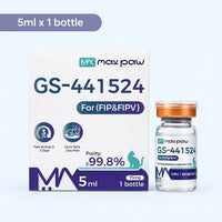
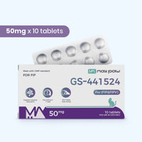
For over fifty years, receiving an FIP diagnosis meant preparing for end-of-life care. Palliative steroids and fluid drainage could buy a cat days or weeks at best. Today, the medical paradigm has completely shifted. FIP is now considered a curable disease thanks to antiviral small molecules, primarily GS-441524 and its related nucleoside analogs.

How GS-441524 Cures FIP
GS-441524 is a small-molecule nucleoside analog that acts as an targeted inhibitor of the viral RNA-dependent RNA polymerase enzyme. When the mutated FIP virus attempts to replicate inside host macrophages, it inadvertently incorporates GS-441524 into its RNA chain. This terminates viral RNA transcription instantly, halting viral replication in its tracks. With viral replication frozen, the cat’s immune system can clear remaining viral loads and repair damaged vascular tissue.
The Standard 84-Day Protocol
The internationally recognized veterinary protocol for FIP antiviral treatment consists of two distinct 84-day phases:
- 84 Days of Active Antiviral Treatment (12 Weeks): The cat receives daily, precisely calculated doses of antiviral medication via subcutaneous injections or oral tablets. Dosing must be strictly maintained every 24 hours without skipping a single day.
- 84 Days of Observation (12 Weeks): Once the 84-day treatment phase ends and blood work normalizes, medication is stopped. The cat enters an 84-day observation window. If no clinical relapse occurs during these 12 weeks, the cat is officially declared cured.
Dosing Considerations by FIP Classification
Dosage calculations vary based on clinical severity and anatomical involvement. Antivirals must cross tissue barriers to reach hidden viral sanctuaries:
- Wet/Effusive FIP: Typically requires the baseline standard dosage (e.g., 5 to 6 mg/kg daily). Response is usually rapid, with fevers breaking in 24 to 48 hours and effusions completely reabsorbing within 7 to 14 days.
- Dry FIP: Requires a slightly higher baseline dosage (e.g., 6 to 8 mg/kg daily) due to dense tissue granulomas that antivirals must penetrate.
- Ocular FIP: Requires increased dosage (e.g., 8 to 10 mg/kg daily) to successfully cross the blood-retinal barrier and resolve ocular lesions.
- Neurological FIP: Requires the highest dosage tiers (e.g., 10 to 12+ mg/kg daily) to cross the tight blood-brain barrier and eliminate viral activity within the central nervous system.
Essential Supportive Care & Home Management
While antiviral drugs target the virus, comprehensive supportive nursing care keeps the patient stable during the critical initial recovery phase:
- Hepatoprotective Supplements: Antiviral processing and rapid cellular breakdown put temporary strain on the liver. Administering liver support compounds like SAMe (S-Adenosylmethionine) and Milk Thistle (Silymarin) helps maintain healthy liver enzymes.
- Nutritional Management: Anorexic cats need immediate nutritional interventions. High-calorie, high-protein wet foods, liquid recovery diets, or temporary placement of a feeding tube (E-tube) prevent hepatic lipidosis.
- Thermoregulation and Comfort: Provide warm heating pads, quiet orthopedic bedding, and minimize environmental commotion to keep stress hormones low.
Can You Prevent FIP? Best Practices for Multi-Cat Homes
Because benign feline coronavirus is ubiquitous across the cat population, eradicating the virus entirely is virtually impossible in multi-cat households or rescues. However, cat parents can take proactive environmental and lifestyle steps to dramatically minimize viral loading and lower mutation triggers:
- Strict Litter Box Sanitation: FCoV spreads through fecal matter. Maintain the gold-standard litter box rule: provide 1 box per cat plus 1 extra. Scoop litter boxes at least twice daily, keep boxes far away from food and water stations, and completely wash and sanitize boxes with diluted bleach or potassium peroxymonosulfate monthly.
- Reduce Environmental Overcrowding: Keep total cat numbers manageable relative to home square footage. Overcrowding increases viral shedding intensity and elevates systemic stress levels.
- Proactive Stress Minimization: When introducing new pets, moving to a new home, or traveling, utilize synthetic feline facial pheromone diffusers (such as Feliway) to calm the nervous system. Schedule routine veterinary procedures (like dental cleanings or spay/neuter operations) only when cats are in peak physical health.
- High-Quality Diet and Immune Support: Feed balanced, protein-rich diets free from unnecessary fillers. Supplementing omega-3 fatty acids (EPA/DHA) and gut-supporting probiotics fosters a healthy microbiome and robust mucosal immunity.